Eye2Brain geometry
We developed from a CAD (Computer Aided Design) drawing a geometry of the whole eye, selecting some appropriate parameters that can be tuned by the user in order to be patient-specific as mush as possible (Fig. 1). For example the thickness of the retina, the depth of the lamina, the diameter of the ocular bulb are some of the main features of the mesh that are clinically relevant and can be modified.
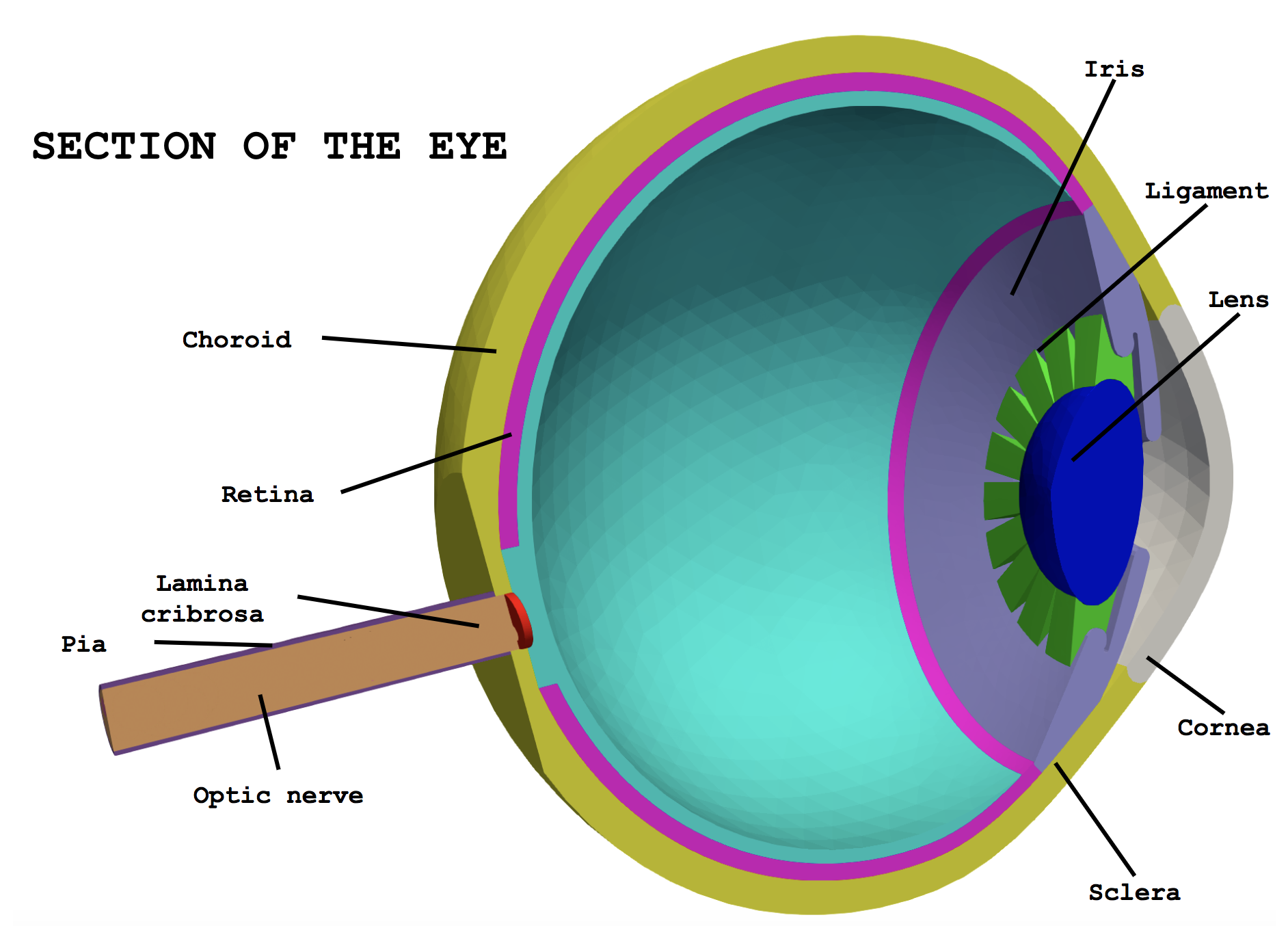
Figure 1: cross section of the ocular geometry
The mesh is structured in order that simulations are available also on single parts of the eye as the whole geometry depending on the application the user needs. Therefore the idea of having a full description of the ocular system is addressed to obtain a virtual laboratory that complies with physical laws. We presented this concept in a poster at the annual meeting of the Association for Research in Visual Ophthalmology (ARVO) [Sala2017arvo].
Bibliography
-
[Sala2017arvo] L. Sala, C. Prud’homme, D. Prada, F. Salerni, C. Trophime, V. Chabannes, M. Szopos, R. Repetto, S. Bertoluzza, R. Sacco, A. Harris, and G. Guidoboni. Patient-specific virtual simulator of tissue perfusion in the lamina cribrosa. In ARVO 2017 Congress. Poster session Imaging: Macula Retina, Blood Flow, OCT Angiography, Baltimore(MD), USA, 07/05/2017.